|
Mutliorganic diagnosis using dental radiographs
|
|
|
|
Osteoporosis, which is a disease that the density of bones becomes low and bones become easily broken, is now an important problem. The osteoporosis is usually diagnosed by the bone density measurement of thighbones. However, a patient who feels no subjective symptom does not usually consult a doctor, and it causes a problem that the osteoporosis cannot be found until a bone break happens. If the osteoporosis can be found by analyzing mandibular radiographs taken at dental clinics and a dentist can recommend the patients to consult a orthopedist, a proper treatment can be applied before the bone break happens.
Professor Akira Taguchi, our co-researcher of Matsumoto Dental University, has found that the reduction of the amount of trabecula, which is a net of bone structure inside a bone, and the thinning and fractuation of cortical bone in a mandible are closely related to the osteoporosis*). Our research extracts trabeculae and cortical bones dental panoramic radiographs, which capture the gnathic bone and teeth from all around the face, and measures the trabecular density and the cortical bone width and strucutre. This is utilized to the support to the diagnosis of osteoporosis and the investigation of the relationship between the osteoprosis and various factors as aging, etc.
It is important to achieve the extraction especially from the panoramic radiographs. The extraction of trabeculae from the image captured by computed radiography (CR), which captures images of very high quality and inputs them directly into computers without using any film or chemical process, has been already archived. However, all dental clinics do not have the CR. The panoramic radiograph system is used in every clinic, and it is easy to collect a large amount of panoramic radiographs. Thus the extraction from the panoramic radiographs helps epidemic researches on the osteoporosis. It is known that dental panoramic radiographs have information on diseases other than osteoporosis, such as arteriosclerosis, and we are developing an automatic multiorganic diagnosis system.
*) "Dental X-rays Could Be First Step In Osteoporosis Screening" (ScienceDaily)
|
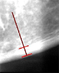
Measurement of cortical bone. Both of the edges of the cortical bone are automatically detected, as shown by the red lines in the figure, and the width is measured, by manually clicking the mental foramen and any point in the cortical bone.
|
We also develop a web-based system for evaluating the effectiveness of osteoporosis diagnosis based on manual observation by dental radiographic experts by collecting the results of diagnosis via the internet.
Selected publications
- S. Takano, M. Muneyasu, S. Yoshida, A. Asano, N. Dewake, N. Yoshinari, and K. Uchida, "Application of Adversarial Training in the Detection of Calcification Regions from Dental Panoramic Radiographs," IEICE Trans. Fundamentals, E108-A, 3, 352-356 (2025. 3).
[

- K. Kohinata, Y. Ishioka, S. Yamada, N. Sugino, H. Kuroiwa, N. Yoshinari, A. Asano, M. Muneyasu, and K. Uchida, "Study on the Carotid Artery Calcification Appearing on the Panoramic Radiography
and Computed Tomography," Journal of Hard Tissue Biology, 28, 1, 93-96 (2019. 1).
- K. Uchida, N. Sugino, S. Yamada, K. Kuroiwa, N. Yoshinari, A. Asano, A. Taguchi, and M. Muneyasu, "Clinical Significance of Carotid Artery Calcification seen on Panoramic Radiographs," Journal of Hard Tissue Biology, 23, 4, 461-466 (2014. 10).
- T. Nakamoto, A. Taguchi, M. Ohtsuka, Y. Suei, M. Fujita, M. Tsuda, M. Sanada, Y. Kudo, A. Asano, and K. Tanimoto, "A computer-aided diagnosis system to screen for osteoporosis using dental panoramic radiographs," Dentomaxillofacial Radiology, 37, 5, 274-281 (2008. 7).
- A. Taguchi, A. Asano, M. Ohtsuka, T. Nakamoto, Y. Suei, M. Tsuda, Y. Kudo, K. Inagaki, T. Noguchi, K. Tanimoto, R. Jacobs, E. Klemetti, S. C. White, K. Horner, OSPD International Collaborative Group, "Observer Performance in Diagnosing Osteoporosis by Dental Panoramic Radiographs: Results from the Osteoporosis Screening Project in Dentistry (OSPD)," BONE, 43, 1, 209-213 (2008. 7).
- Agus Zainal Arifin, A. Asano, A. Taguchi, T. Nakamoto, M. Ohtsuka, M. Tsuda, Y. Kudo, and K. Tanimoto, "Use of Fuzzy Neural Network in Diagnosing Postmenopausal Women with Osteoporosis Based on Dental Panoramic Radiographs," Journal of Advanced Computational Intelligence and Intelligent Informatics, 11, 8, 1049-1058 (2007. 10).
- Febriliyan Samopa, A. Asano, and A. Taguchi, "Automatic Region-of-Interest Extraction from Dental Panoramic Radiographs for Forensic Personal Identification," Proc. 2nd Korean-Japan Joint
Workshop on Pattern Recognition (KJPR2007), (IEICE Tech. Rep. 107, 281) 161-166 (2007. 10).
- Agus Zainal Arifin and A. Asano, "Image Segmentation by Histogram Thresholding Using Hierarchical Cluster Analysis," Pattern Recognition Letters, 27, 13, 1515-1521 (2006. 10).
- A. Asano, T. Tambe, A. Taguchi, C. Muraki Asano, T. Nakamoto, K. Tanimoto, M. Muneyasu, and T. Hinamoto, "Extraction of trabecular structures of mandible excluding tooth roots on dental panoramic radiographs using mathematical morphology," Proc. 18th International Conference on Pattern Recognition (ICPR2006), 3, 988-991 (2006. 8).
- Agus Zainal Arifin, A. Asano, A. Taguchi, T. Nakamoto, M. Ohtsuka, M. Tsuda, Y. Kudo, and K. Tanimoto, "Computer-aided system for measuring the mandibular cortical width on dental panoramic radiographs in identifying postmenopausal women with low bone mineral density," Osteoporosis International, 17, 5, 753-759 (2006. 5) (Online issue DOI: 10.1007/s00198-005-0045-2, 2006. 3). [corresponding author]
- A. Asano, A. Taguchi, T. Nakamoto, K. Tanimoto, and Agus Zainal Arifin, "Apparatus for assisting diagnosis of osteoporosis," Patent pending in Japan, 2006-213259, 2008-036068 (applied on Aug. 4, 2004, and disclosed on Feb. 21, 2008).
- A. Asano, A. Taguchi, T. Nakamoto, K. Tanimoto, and Agus Zainal Arifin, "Apparatus for assisting diagnosis of osteoporosis," Patent PCT-disclosed, 2004-304855, PCT/JP2005/019078, WO/2006/043523 (applied in Japan on Oct. 20, 2004, PCT-applied on Oct. 18, 2005, and PCT-disclosed on Apr. 27, 2006).
- A. Taguchi, T. Nakamoto, and A. Asano, "Osteoporosis CAD using panoramic radiograph," Patent in Japan, No. 3964795 (applied on Jan. 7, 2003 [2003-001395], disclosed on Jul. 29, 2004 [2004-209089], and fixed on Jun. 1, 2007).
Other works in the field of medical image processing:
- Febriliyan Samopa, A. Asano, and A. Taguchi, "Automatic Region-of-Interest Extraction from Dental Panoramic
Radiographs for Forensic Personal Identification," Proc. 2nd Korean-Japan Joint
Workshop on Pattern Recognition (KJPR2007), (IEICE Tech. Rep. 107, 281) 161-166 (2007. 10).
- A. Asano, S. Hayashi, M. Muneyasu, M. Furumura, and J. Nakayama, "Application of morphological size distribution analysis to the evaluation of anti-aging treatments for skin rejuvenation," Proc. Joint 3rd International Conference on Soft Computing and Intelligent Systems and 7th International Symposium on advanced Intelligent Systems (SCIS & ISIS 2006), 1784-1788 (2006. 9).
- S. Nakamura, A. Yoshida, A. Asano, and T. Hinamoto, "Radiograph Simulation System Using a Virtual Head Phantom," Meeting Abstracts of 90th Scientific Assembly and Annual Meeting, the Radiological Society of North America (RSNA04), 814 (2004. 11).
|